

Our Medical Image Registration Features
With Chimaera's Image Registration, it becomes possible to compensate for misalignment up to and including complex patient movements between different image acquisitions, whether for follow-up images, stitching of images, or acquisitions with different imaging modalities such as CT, MR or PET/SPECT.
Provided as an OEM component with C-API, our Image Registration is perfect for extending existing customer applications with additional features that require accurate spatial mapping.
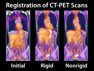
Rigid Registration
- Rotation and translation only
- Image content is not modified
- No manual parameterization needed
- 2D, 3D, and time series
- Supports multi-modal data (e.g. CT, MR, PET, SPECT, etc)
Nonrigid Registration
- Computes complex deformations between images
- Deformation field can compensate for respiratory motion, cardiac motion, patient positioning, etc
- Easy and flexible parameterization for different use cases
- 2D, 3D, and time series
- Supports multi-modal data (CT, MR, PET, SPECT, etc)
Applications
- Retrospective image fusion
- Follow-up studys
- Difference and perfusion imaging
- 4-D motion correction
- Model / atlas-to-image registration
- Post market surveillance
Solutions
- Rigid image registration
- Nonrigid image registration
- Support of 2-D & 3-D image data
Benefits
- Simple integration (C-API)
- Low hardware requirements (CPU based)
- Fully automatic rigid registration
- No / minimal parameterisation (nonrigid)
- Quality documents for product integration
Applications for Medical Image Registration
Rigid Registration
The transformation between the images is limited to only rotations and translations. This method is often used to establish a link between different scanner coordinate systems or to account for rigid body movements while the image content does not change:
- Retrospective image fusion (e.g. for PET/CT, PET/MR, etc)
- Side-by-side viewing of follow-up studies
- Image stitching
The following example shows 3D overlay visualizations of CT and SPECT brain scans of the same patient acquired separately (no hybrid scanner).

This image shows the initial state without registration.

This image shows the corrected image position as computed by our rigid registration.
Nonrigid Registration
Nonrigid (also sometimes denoted as deformable or elastic) image registration techniques allow far more degrees of freedom in terms of deformation compared to rigid registration. Complex deformations between images often need to be calculated to compensate for respiratory motion, cardiac motion, slight differences in patient positioning, or other types of soft tissue misalignments. Cross-patient registration is also possible to create atlases on different subjects.
- Perfusion imaging
- Difference imaging
- Atlas registration
- Transfer findings to follow-up study
In the following use case, we demonstrate how nonrigid image registration can compensate complex organ movement between a non-contrasted and an arterial phase of an MR perfusion image study.

This is the initial 3D relation of the two MR series shown in a coronal plane that crossfades between the two phases. For perfusion calculations, it is necessary to compensate for organ motion between acquisitions.

As a result, after applying the deformation calculated with our nonrigid image registration, the organ motion is now corrected. This provides a good basis for calculations, especially for the liver application shown in this example.

With the so-called checkerboard visualization shown above, blocks of images are displayed alternately. This enables an assessment of how well structures fit together before and after registration.

The image above shows the warping of the arterial MR series along the deformation field computed with our nonrigid image registration.
Integration as OEM Component
No worries about integrating our technically demanding solutions - we make it easy for our customers!
Our Image Registration is available as a dynamic library with an application programming interface (written in C), with no additional dependencies required. The integration into customer applications is very simple due to the minimal parameterization (only required for the non-rigid registration) and existing code examples.
And with regard to certification, the use of our registration is also very simple thanks to the corresponding quality documents.
- Single dynamic library with C-API
- Simple integration into customer code
- Example interface code with wrappers for ITK image types
- Output as transformation or deformation vector field in addition to resampled output images
- Quality documentation for fast integration into certified medical software
Did you know that our Chimaera SDK brings your project from prototype to finished product?

Volker

Dr.-Ing. Volker Daum
Am Weichselgarten 7
91058 Erlangen
Germany
+49 (0)9131 - 691 385
+49 (0)9131 - 691 386
Dr. Volker Daum, Chimaera CTO