

Einführung
Bei der modernen Operationsplanung von Hüfte, Becken oder Oberschenkel spielt die CAD-Modellierung eine wesentliche Rolle. Dies ist vor allem bei maßgefertigten Implantaten der Fall, die genau auf die Anatomie des Patienten zugeschnitten sind.
Bevor mit der patientenbasierten CAD-Bearbeitung begonnen werden kann, muss zunächst das Modell erstellt werden. Zu diesem Zweck werden die anatomischen Zielstrukturen (Hüfte, Becken usw.) aus den CT-Bildern des Patienten segmentiert, wobei standardmäßig halbautomatische Softwaretools zum Einsatz kommen.
Insbesondere bei individuellen anatomischen oder pathologischen Gegebenheiten wie vorbestehenden Implantaten, verschiedenen Frakturen oder einem Osteosarkom stoßen bestehende Brush-Annotation-Tools oder schwellenwertbasierte Segmentierungen an ihre Grenzen.
Dies macht diesen Vorverarbeitungsschritt zeit- und kostenaufwendig.
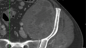
Die von Krebs umgebene linke Hüftkugel wurde manuell und pixelgenau mit dem Chimaera Smart Brush annotiert.
Dieser Blogbeitrag zeigt anhand des Fallbeispiels der automatischen Segmentierung von Hüfte und Oberschenkelknochen, wie der Weg zur Erstellung eines KI-Modells verläuft und welche Voraussetzungen dafür erfüllt sein müssen.
Durch präzise Ingenieursarbeit kann ein solches vollautomatisches KI-Modell in den Arbeitsablauf des Kunden integriert werden. Es führt in Sekundenschnelle eine vollständige Segmentierung des Beckens, des Oberschenkels und der Implantate durch.
Methoden und Workflow
There is very rarely the ideal out-of-the-box AI model that is perfectly suited to the problem at hand.
Several steps are necessary before an AI model can automate the target task of segmenting pelvic blades and femurs in the daily work process.
First, the algorithm is specifically tailored to the customer's problem (which anatomical regions, which implants, specific products or manufacturers, etc.). Second, due to data protection and customer agreements, an AI model trained on client A's database cannot be distributed to client B.

Arbeitsschritte für die Erstellung eines KI-Modells.
Der Vorgang, bis das KI-Modell auf Knopfdruck Segmentierungen für den Kunden generiert, lässt sich grundsätzlich in vier Stufen unterteilen.
Grundsätzlich wird ein geeigneter Bestand an CT-Bildern und zugehörigen Datenannotationen (manuelle Segmentierungen) benötigt. Auf dieser Basis erfolgt das eigentliche Engineering, wobei dem Algorithmus iterativ beigebracht wird, die Aufgabe auf Basis des vorhandenen Datenbestandes zu erlernen, um mit neuen Bilddaten eine vollautomatische Segmentierung durchzuführen. Ziel der Integrationsphase ist es, das KI-Modell so vorzubereiten, dass es nahtlos in die Verarbeitungsprozesse des Kunden integriert werden kann.
Bei Chimaera legen wir Wert darauf, keine Blackbox-Lösungen zu verkaufen: Durch Schulungen, Quellcode- und Know-how-Transfer befähigen wir unsere Kunden, eigenständig zu entwickeln und zukünftige Anpassungen umzusetzen.
Alle diese Schritte werden durch die Chimaera-Dienstleistungen aus einer Hand erbracht und können so optimal aufeinander abgestimmt werden.
Datenmanagement
In einem ersten Schritt sollte die genaue Aufgabenstellung im engen Austausch mit dem Kunden definiert werden. In diesem Beispiel ist die automatische Segmentierung von 4 Klassen gefordert:
- linke Hüftpfanne
- rechte Hüftpfanne
- linker Oberschenkelknochen
- rechter Oberschenkelknochen
Würde man die Aufgabe auf Fälle von Hüftrevisionen ausweiten, müssten auch die vorhandenen Implantate segmentiert und zusätzliche Klassen definiert werden. In diesem Fall wäre es denkbar, diese als eine universelle Klasse (z.B. Implantat) beizubehalten oder eine genauere Unterteilung vorzunehmen (z.B. Pfanne, Schaft, Schrauben, etc.) - je nach Anwendung und Vorstellung des Kunden.
Annotation
Für jeden einzelnen Datensatz ist eine vollständige Annotation der definierten Klassen erforderlich. Diese dient der KI in der Trainingsphase als Input und ermöglicht es ihr, Formen und Strukturen in den CT-Scans zu erkennen.
Da der KI-Algorithmus die pixelgenauen Markierungen als Ground Truth interpretiert, sind qualitativ hochwertige Annotationen unerlässlich. Die Ergebnisse der endgültigen KI-Lösung sind nur so gut wie die Vorarbeit bei den Annotationen.
Oft können auch die vom Kunden über Jahre hinweg durchgeführten Segmentierungen als Annotationen dienen. Sollte dies nicht oder nur unzureichend der Fall sein, bietet Chimaera die Annotation als Dienstleistung durch unser medizinisch geschultes Team an. Die hohe Qualität wird durch den Einsatz unserer speziell entwickelten Software und den integrierten Qualitätsprozess gewährleistet.
Im Allgemeinen gilt: Je komplexer die Aufgabe und je höher die gewünschte Genauigkeit, desto mehr Daten werden für das Training des KI-Modells benötigt. Die benötigte Datenmenge kann dabei zwischen 100 und 10.000 Datensätzen liegen. Außerdem ist darauf zu achten, dass die variablen Parameter wie Geschlecht, Bevölkerung, Pathologien, Implantate usw. möglichst breit gefächert sind, damit das KI-Modell möglichst generische Regeln lernen kann.
KI Engineering
Sobald erste Daten und Annotationen verfügbar sind, kann ein KI-Prototyp trainiert werden.
Dieser wird noch keine perfekten Segmentierungen liefern, aber es lassen sich Tendenzen für weitere Optimierungen erkennen, beispielsweise in Bezug auf die Netzwerkarchitektur, das Preprocessing oder Fehler in der Annotation. Darüber hinaus kann im engen Austausch mit dem Kunden und seinem Anwendungs-Know-how an den Feinheiten gearbeitet werden.
Auch danach bleibt die Erstellung des KI-Modells ein interaktiver Prozess, bis schließlich der endgültige Algorithmus entsteht. Selbst hier können regulatorische Anforderungen eine ständige Überprüfung und Verbesserung erfordern.
Bei Bedarf (z. B. Datenschutz) können alle Schulungen auch direkt beim Kunden vor Ort durchgeführt werden.
Integration und Implementierung
Liefert die KI die Segmentierungen von Hüfte und Oberschenkel in der gewünschten Qualität, kann der Algorithmus in die Arbeitsabläufe des Kunden integriert werden.
In seiner einfachsten Form läuft dieser Verarbeitungsschritt als eigenständiger Prozess auf dem Arbeitsrechner ab, der CT-Datensätze einliest und die fertige Segmentierung an einem definierten Pfad ablegt.
Alternativ kann das KI-Modell auch direkt in eine bestehende Softwarelösung integriert werden.
Unabhängig von der Art der Integration stellt Chimaera dem Kunden den vollständigen Quellcode des KI-Modells zur Verfügung. Ebenso wird das Know-how der neuen Technologie an die Mitarbeiter weitergegeben, sodass eigenständige Weiterentwicklungen des Algorithmus möglich sind.
Ergebnis
Je nach Komplexität kann so innerhalb weniger Arbeitswochen ein präzises KI-Modell entwickelt werden, das – wie im Beispiel – Hüftpfannen und Oberschenkelknochen vollautomatisch segmentiert.
Das Modell kann zu einem späteren Zeitpunkt zusätzlich auf neue Krankheitsbilder trainiert werden. So könnten im Falle der Implantaterkennung neue Hersteller oder Modelle problemlos hinzugefügt werden.
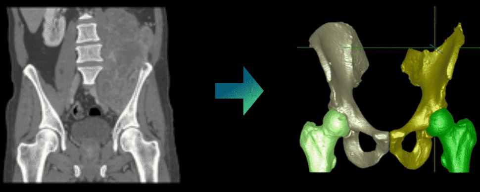
Das fertige KI-Modell kann anschließend verwendet werden, um anatomische Strukturen aus einer 3D-CT-Serie vollautomatisch zu segmentieren.