Use case: Compensation of Cardiac Motion in 3D MR Time Series
This use case demonstrates the compensation of cardiac motion in a 3D time series acquired with an MR scanner. The reconstructed single 3D images were reconstructed at specific phases of the cardiac cycle using cardiac gating.
Please see our page on Image Registration for more details regarding the component.
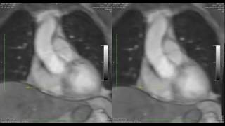
The images show the same coronal slice through the 3D MR image with the original time series on the left side and the time series after correcting cardiac motion using our Image Registration on the right.

Precise motion compensation thanks to non-rigid image registration
In the example shown, a specific anatomical landmark is marked with an arrow to illustrate movement through the cardiac cycle.
For certain applications, such as perfusion measurements within the anatomy of the heart, it is desirable to "freeze" the cardiac motion to a specific point within the cardiac cycle without altering the image intensities in a way that compromises the measurements.
As shown in the example, this is achieved by compensating for heart motion using our non-rigid image registration technique. As can be seen in the resampled time series on the right, the orientation arrow remains at the desired anatomical position throughout.
